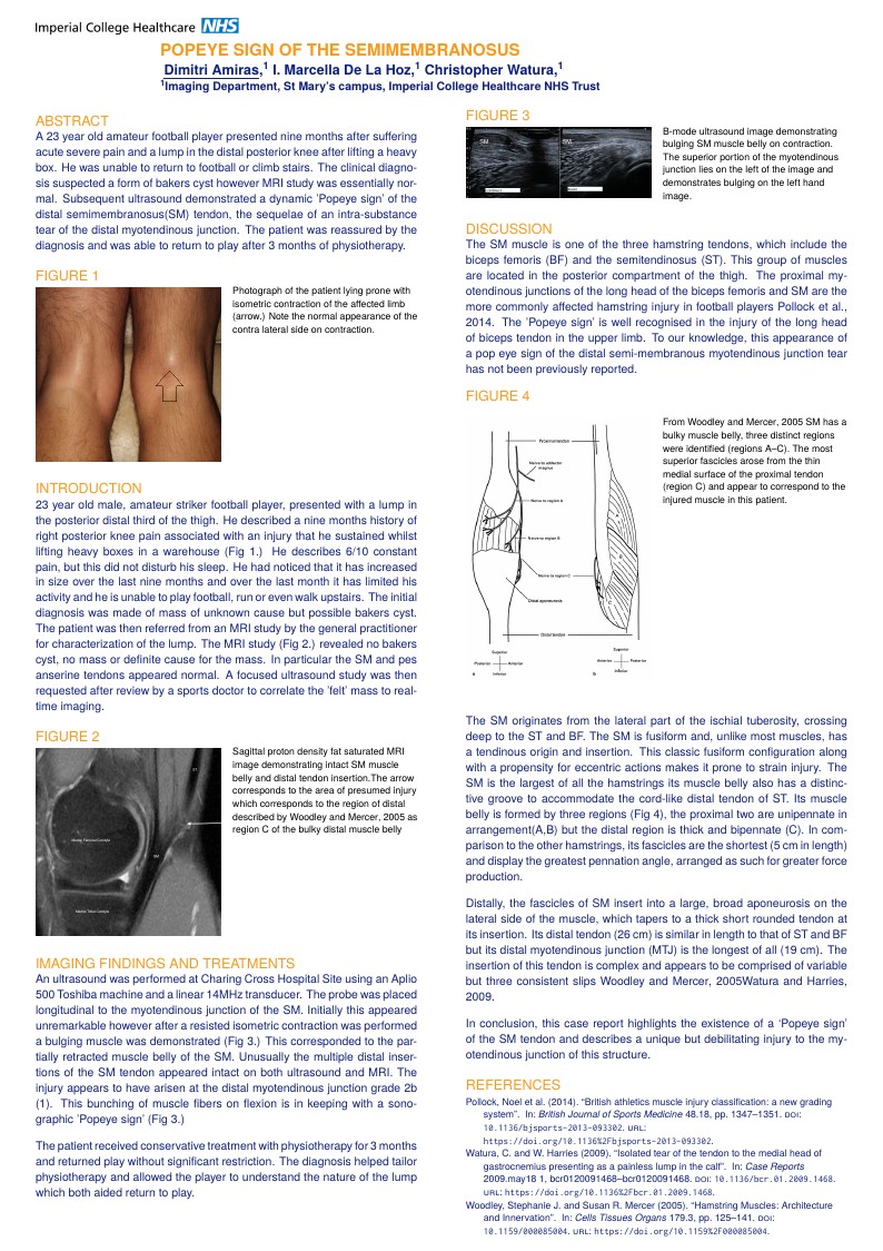
Popeye sign of the semimembranosus
Auteur:
Dimitri Amiras
Last Updated:
il y a 9 ans
License:
Creative Commons CC BY 4.0
Résumé:
Case report poster

\begin
Discover why over 25 million people worldwide trust Overleaf with their work.
Case report poster

\begin
Discover why over 25 million people worldwide trust Overleaf with their work.
%% beamerthemeImperialPoster v1.0 2016/10/01
%% Beamer poster theme created for Imperial College by LianTze Lim (Overleaf)
%% LICENSE: LPPL 1.3
%%
%% This is the example poster demonstrating use
%% of the Imperial College Beamer Poster Theme
\documentclass[xcolor={table}]{beamer}
%% Possible paper sizes: a0, a0b, a1, a2, a3, a4 (although Imperial College posters are usually A0 or A1).
%% Possible orientations: portrait, landscape
%% Font sizes can be changed using the scale option.
\usepackage[size=a0,orientation=portrait,scale=1.4]{beamerposter}
\usepackage{ragged2e}
\usetheme{ImperialPoster}

%% Four available colour themes
\usecolortheme{ImperialWhite} % Default
% \usecolortheme{ImperialLightBlue}
% \usecolortheme{ImperialDarkBlue}
% \usecolortheme{ImperialBlack}

\title{Popeye sign of the semimembranosus}

\author{\ \mainauthor{Dimitri Amiras},\Tsup{1} I. Marcella De La Hoz,\Tsup{1} Christopher Watura,\Tsup{1} }

\institute{\Tsup{1}Imaging Department, St Mary's campus, Imperial College Healthcare NHS Trust}

\addbibresource{biblio.bib}


\begin{document}
\begin{frame}[fragile=singleslide,t]\centering

\maketitle
\begin{columns}[onlytextwidth,T]

%%%% First Column
\begin{column}{.47\textwidth}


\begin{block}{Abstract}
\justifying
A 23 year old amateur football player presented nine months after suffering acute severe pain and a lump in the distal posterior knee after lifting a heavy box. He was unable to return to football or climb stairs. The clinical diagnosis suspected a form of bakers cyst however MRI study was essentially normal. Subsequent ultrasound demonstrated a dynamic 'Popeye sign' of the distal semimembranosus(SM) tendon, the sequelae of an intra-substance tear of the distal myotendinous junction. The patient was reassured by the diagnosis and was able to return to play after 3 months of physiotherapy.
\end{block}


\begin{sidefigure}

\includegraphics[width=\hsize]{photocontractarrow.png}
\caption{Photograph of the patient lying prone with isometric contraction of the affected limb (arrow.) Note the normal appearance of the contra lateral side on contraction.

}
\end{sidefigure}

\begin{block}{Introduction}
\justifying
23 year old male, amateur striker football player, presented with a lump in the posterior distal third of the thigh.
He described a nine months history of right posterior knee pain associated with an injury that he sustained whilst lifting heavy boxes in a warehouse (Fig 1.) He describes 6/10 constant pain, but this did not disturb his sleep. He had noticed that it has increased in size over the last nine months and over the last month it has limited his activity and he is unable to play football, run or even walk upstairs. The initial diagnosis was made of mass of unknown cause but possible bakers cyst. The patient was then referred from an MRI study by the general practitioner for characterization of the lump.
The MRI study (Fig 2.) revealed no bakers cyst, no mass or definite cause for the mass. In particular the SM and pes anserine tendons appeared normal. A focused ultrasound study was then requested after review by a sports doctor to correlate the 'felt' mass to real-time imaging.
\end{block}
\begin{sidefigure}
\includegraphics[width=\hsize]{MRI.png}
\caption{Sagittal proton density fat saturated MRI image demonstrating intact SM muscle belly and distal tendon insertion.The arrow corresponds to the area of presumed injury which corresponds to the region of distal described by \cite{Woodley_2005} as region C of the bulky distal muscle belly}
\end{sidefigure}

\begin{block}{Imaging Findings and Treatments}
\justifying
An ultrasound was performed at Charing Cross Hospital Site using an Aplio 500 Toshiba machine and a linear 14MHz transducer. The probe was placed longitudinal to the myotendinous junction of the SM. Initially this appeared unremarkable however after a resisted isometric contraction was performed a bulging muscle was demonstrated (Fig 3.) This corresponded to the partially retracted muscle belly of the SM. Unusually the multiple distal insertions of the SM tendon appeared intact on both ultrasound and MRI. The injury appears to have arisen at the distal myotendinous junction grade 2b (1). This bunching of muscle fibers on flexion is in keeping with a sonographic 'Popeye sign' (Fig 3.)

The patient received conservative treatment with physiotherapy for 3 months and returned play without significant restriction. The diagnosis helped tailor physiotherapy and allowed the player to understand the nature of the lump which both aided return to play.
\end{block}
\end{column}

%%%% Second Column
\begin{column}{.47\textwidth}
\begin{sidefigure}
\includegraphics[width=\hsize]{us_RELAX+CONTRACT.png}
\caption{B-mode ultrasound image demonstrating bulging SM muscle belly on contraction. The superior portion of the myotendinous junction lies on the left of the image and demonstrates bulging on the left hand image.

}
\end{sidefigure}
\begin{block}{Discussion}
\justifying
The SM muscle is one of the three hamstring tendons, which include the biceps femoris (BF) and the semitendinosus (ST). This group of muscles are located in the posterior compartment of the thigh. The proximal myotendinous junctions of the long head of the biceps femoris and SM are the more commonly affected hamstring injury in football players \cite{Pollock_2014}. The 'Popeye sign' is well recognised in the injury of the long head of biceps tendon in the upper limb. To our knowledge, this appearance of a pop eye sign of the distal semi-membranous myotendinous junction tear has not been previously reported.

\begin{sidefigure}
\includegraphics[width=\hsize]{SEMIDRAW.png}
\caption{From \cite{Woodley_2005} SM has a bulky muscle belly, three distinct regions were identified (regions A–C). The most superior fascicles arose from the thin medial surface of the proximal tendon (region C) and appear to correspond to the injured muscle in this patient.
}
\end{sidefigure}
The SM originates from the lateral part of the ischial tuberosity, crossing deep to the ST and BF. The SM is fusiform and, unlike most muscles, has a tendinous origin and insertion. This classic fusiform configuration along with a propensity for eccentric actions makes it prone to strain injury. The SM is the largest of all the hamstrings its muscle belly also has a distinctive groove to accommodate the cord-like distal tendon of ST.  Its muscle belly is formed by three regions (Fig 4), the proximal two are unipennate in arrangement(A,B) but the distal region is thick and bipennate (C). In comparison to the other hamstrings, its fascicles are the shortest (5 cm in length) and display the greatest pennation angle, arranged as such for greater force production.

Distally, the fascicles of SM insert into a large, broad aponeurosis on the lateral side of the muscle, which tapers to a thick short rounded tendon at its insertion. Its distal tendon (26 cm) is similar in length to that of ST and BF but its distal myotendinous junction (MTJ) is the longest of all (19 cm). The insertion of this tendon is complex and appears to be comprised of variable but three consistent slips \cite{Woodley_2005}\cite{Watura_2009}.

In conclusion, this case report highlights the existence of a ‘Popeye sign' of the SM tendon and describes a unique but debilitating injury to the myotendinous junction of this structure.

\end{block}

\printbibliography

\end{column}
\end{columns}


\end{frame}
\end{document}